
Project 2: Efficient Mapping of Arterial Spin Labeling Using 3D GRASE Imaging
Project Leader: David Feinberg, M.D, Ph.D; (Advanced MRI Technologies, LLC)
The overall goal of this project is to develop new methods for quantitative measurements of cerebral blood flow (CBF) and arterial transit time (ATT), also called bolus arrival time (BAT) in arterial spin labeling perfusion studies for specific applications to support the research projects and clinical applications of the Resource Center. Arterial spin labeling (ASL) MRI has great promise for quantitative measurements of brain function, because the method is entirely noninvasive and can be repeated rapidly and in principle indefinitely. However, due to the small volume fraction (3- 5%) of blood vessels compared to brain tissue, ASL-MRI yields an intrinsically small signal and consequently poor SNR.
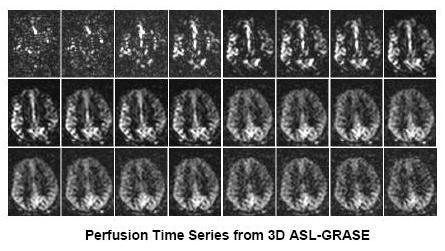
Recently, 3D imaging of the ASL signal has become possible, providing higher SNR efficiency than conventional 2D techniques, because data acquired at every step for image formation contributes to the ASL signal. Furthermore, 3D ASL imaging facilitates measuring the time course of the ASL signal simultaneous throughout the brain, as there is identical inflow time in all segmented 3D slices, unlike multi-slice 2D techniques. However, the development of 3D ALS techniques has been complicated by the demand for fast single-shot acquisitions while avoiding geometrical distortions and signal loss. Over the past decade, the investigator of this project has developed new 3D acquisition methods resulting in higher efficiency, sensitivity, and precision. This application to create new cycled labeling methods represents new efforts to develop improved acquisition techniques focused on the detection of neurodegenerative diseases.